Earlier this year, the journal Bulletin of the History of Medicine published a selection of papers called “Beyond Illustrations: Doing Anatomy with Images and Objects.” The articles examine the importance and impact of the visualization of anatomy, pathology, and disease. Carin Berkowitz, director of the Beckman Center for the History of Chemistry at the Chemical Heritage Foundation, guest edited the forum and joined us for a Q&A.
This forum has been a long time in the making. How happy are you to see it come to life?
I am thrilled to see the issue out! I think Eva Ahren and I first realized that there were a number of people working on visualization and anatomy at the annual meeting of the American Association for the History of Medicine in 2011 and began to think about the special issue then, so it has been four years in the making.

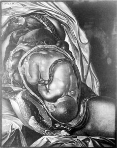
Mezzotint by Johann Michael Seligmann from a drawing by Jan Van Rymsdyk, for Charles Nicholas Jenty’s The Demonstrations of a Pregnant Uterus of a Woman at Her Full Time: In Six Tables, as Large as Nature (first German edition, Nuremberg, 1761, from first London edition, 1757). Broadsheet (627 × 443 mm). Courtesy of the Wellcome Library, London. From Carin Berkowitz’s article “Authorship, Patronage, and Illustrative Style in Anatomy Folios, 1700–1840.”
How helpful is it for a journal like the Bulletin to allow space for something like this?
I think it is quite remarkable and very important for a journal like Bulletin to include a forum like this. The journal’s editors, Mary Fissell and Randall Packard, were very supportive of our efforts, and perhaps most importantly, no one at the journal ever told us that we needed to limit the number of illustrations in the issue. As you might imagine, figures and images are essential to articles that attempt to address issues of visual and material culture, and yet they are tricky and expensive to print and therefore often restricted. My article in the issue depends on the comparison of what would normally be considered a huge number of illustrations; to give you a sense of how generous Bulletin was, in fact, there are almost as many pictures in my short article as there are in my entire forthcoming book. Nico Bertoloni Meli’s piece depends on large numbers of images as well. I think we were all surprised and very grateful that the journal gave us the opportunity to address the intersections of medicine, visual culture, and material culture properly.
What is the importance of viewing illustrations beyond that context and using them to learn about history and context?
I think that historians sometimes use pictures as decorative objects—the way that you might add a frieze to a building. What we tried to do in this issue is to think about the pictures as the primary object of study; we made them structural to our arguments. By doing that, one can see that images and objects have often shaped ideas, rather than the reverse. Meli’s article shows that the discipline of pathology really arose through and hand-in-hand with, not only the creation of specimens, but also with conventions of representation that allowed disease to be visualized. Lisa O’Sullivan and Ross L. Jones show us how a material specimen could generate not only knowledge, data, and practices, but also whole networks of practitioners and even institutions around it. Both cases offer instructive examples of the historiographical rewards that can be reaped by placing the visual and material at the center of inquiry.

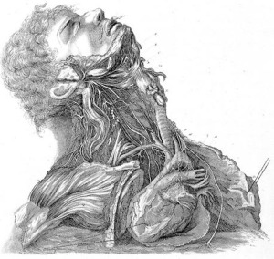
Engraving by J. Grant from a drawing by Charles Bell, Plate II: “Nerves of the Neck” from Charles Bell’s A Series of Engravings Explaining the Course of the Nerves (1803). Quarto (29 cm high). Courtesy of the Wellcome Library, London. From Carin Berkowitz’s article “Authorship, Patronage, and Illustrative Style in Anatomy Folios, 1700–1840.”
How has technology changed the landscape of interacting with visual displays of anatomy?
This is a good question. I won’t try to answer it with respect to modern displays, but with respect to historical ones, I fear sometimes that although it makes visual displays more readily available, it does so in a way that further decontextualizes them. We make a point in our Forum of trying to follow art historians’ conventions (rather than those of historians of science) in labelling, describing, where possible, not only the text from which an image came and that text’s author, but also the makers of the image (artists, engravers), and, importantly, the medium and size within which it was originally produced. This seems to me absolutely essential, and something that often gets stripped away even with reprinting, but all the more so with reproductions available on websites and through apps. When one loses sight of format, one fails to appreciate the possible conditions of use. Some historians, for example, have written about William Hunter’s Anatomy of the Human Gravid Uterus as an instructional text for practitioners, but Hunter’s original book was an elephant folio (the name tells you something about its size). A practitioner could never have used it as a ready reference, as it was hardly portable (or affordable)! Technology hasn’t introduced these problems of decontextualized reproduction, and it offers to solve other problems, like those of reproducing color for journal articles, for example, but image galleries do seem to flatten differences that are rather stark in the material objects themselves.
Where does the conversation go from this point?
Well, there’s much interesting work being done on other kinds of visual displays and material representations within medicine–on models, on photographs, on standardized charts of anatomy. I, for one, would like to see much more intersection between these sorts of conversations and those taking place in art history and visual studies, and even between history of medicine and history of science. It seems to me that there is much that communities gain from interacting, including new vocabularies and new kinds of questions. The history of medicine offers so much under-explored and fertile empirical material that there are many possibilities for future directions!
